surgeonring

The anatomy journal

The body is a living story.

Surgeonring explains anatomy, physiology, biology, and histology as one connected subject — every organ traced from its position in the body down to the cells that make it work, and the emergencies where that knowledge has to be used in minutes.

40 illustrated guidesCited from standard textsWritten to be re-read

Where to start

The journal is written to be read in either direction. Build up from the tissues, or start with an organ you already care about and follow it down.

  1. 01Body systemsTen systems, each a set of organs solving one problem together — and the map of which organ belongs where.
  2. 02Organ guidesEvery major organ traced from its position in the body down to the cells that make it work.
  3. 03Tissues and building blocksThe layer underneath the organs: the four basic tissues, the cells they are built from, and how to tell them apart on a slide.
  4. 04Emergency medicineThe time-critical conditions: how to recognise them, why minutes change the outcome, and what to do before help arrives.

Body systems / 10 studies

Zoom out.
See the whole system.

Ten systems, each a set of organs solving one problem together — and the map of which organ belongs where.

01 / 10
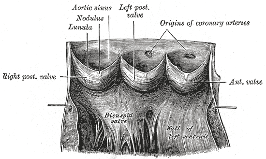
The aortic valve opened out, with the sinuses of Valsalva and the origins of the coronary arteries.

Cardiovascular system / 10 min read

Cardiovascular

One pump, two circuits, and 100,000 kilometres of tubing. How the cardiovascular system moves five litres of blood a minute, what sets blood pressure, and why the whole design is built around a single number.

Open study
02 / 10
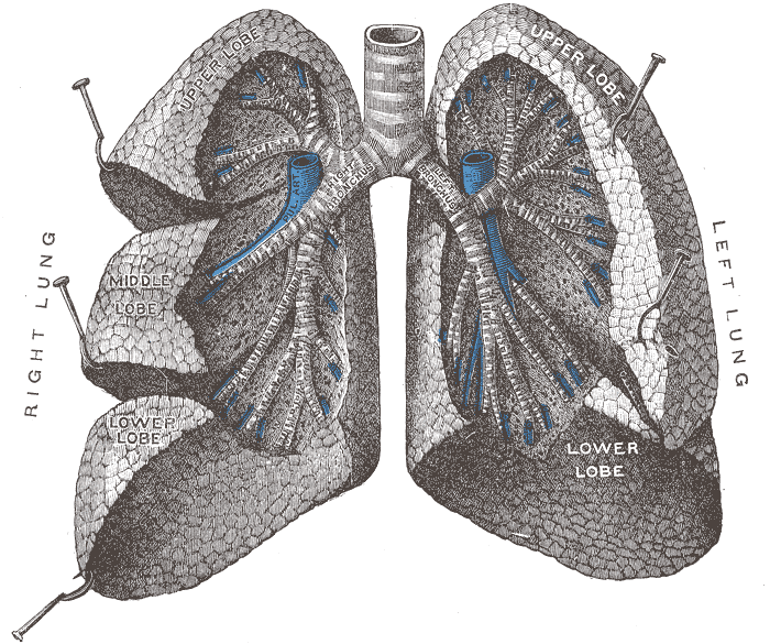
The bronchi and bronchioles, with the lungs drawn apart to expose the branching airway.

Respiratory system / 10 min read

Respiratory

From nose to alveolus in about twenty-three branches. How breathing actually works, why the diaphragm does most of it, how gas crosses a membrane thinner than a soap film, and what controls the rate.

Open study
03 / 10
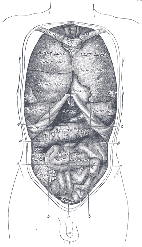
The thoracic and abdominal viscera from the front, in their natural relations.

Digestive system / 10 min read

Digestive

A nine-metre tube, four accessory organs, and a surface area the size of a studio flat. What happens to food at each station, which organ contributes what, and why most absorption is finished before the colon.

Open study
04 / 10
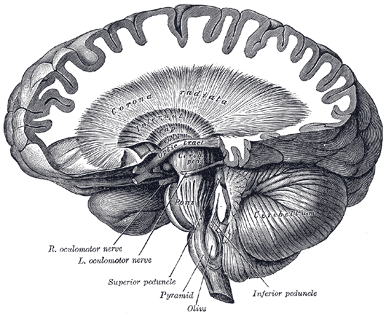
Dissection showing the course of the cerebrospinal fibres from cortex to spinal cord.

Nervous system / 10 min read

Nervous

Eighty-six billion neurons organised into a system that senses, decides and acts in milliseconds. How the central, peripheral and autonomic divisions fit together, and what each one actually controls.

Open study
05 / 10
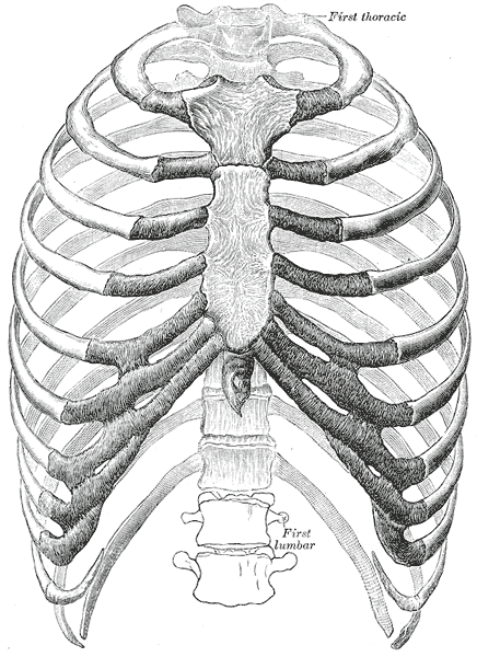
The thorax from the front: ribs, costal cartilages and sternum as one articulated cage.

Skeletal system / 9 min read

Skeletal

206 bones doing five jobs at once — support, protection, movement, mineral storage and blood cell production. How the skeleton is organised, how bone stays alive, and why it is rebuilt continuously.

Open study
06 / 10
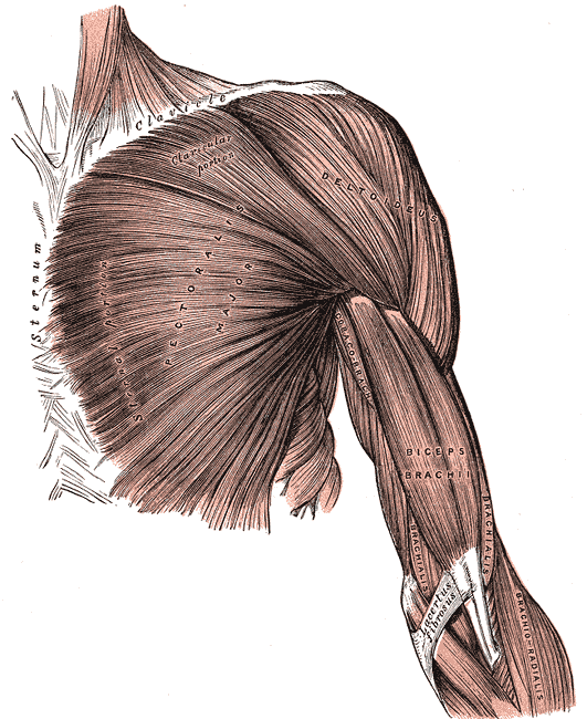
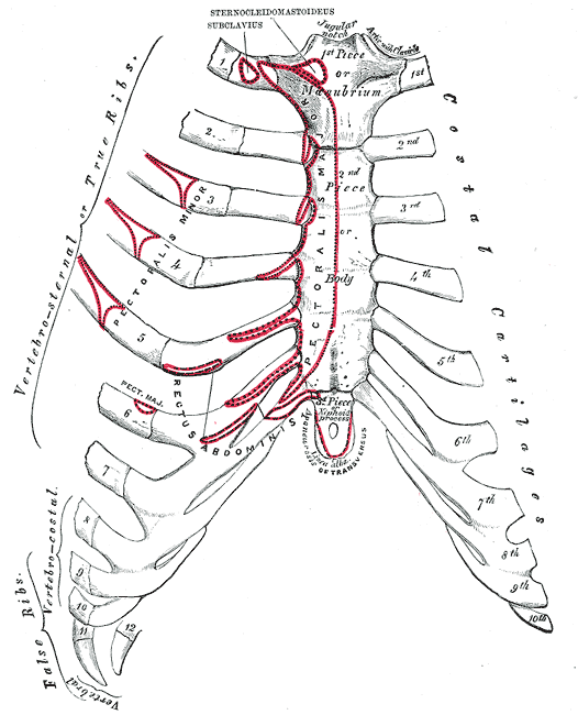
Pectoralis major, showing how a muscle runs from a broad origin to a single tendon.

Muscular system / 10 min read

Muscular

Around 600 skeletal muscles, all pulling and never pushing. How a muscle produces graded force, why they work in opposing pairs, what makes one fibre fast and another slow, and where the energy comes from.

Open study
See all 10 system guides

Organ guides / 15 studies

Choose an organ.
Follow the system.

Every major organ traced from its position in the body down to the cells that make it work.
01 / 15
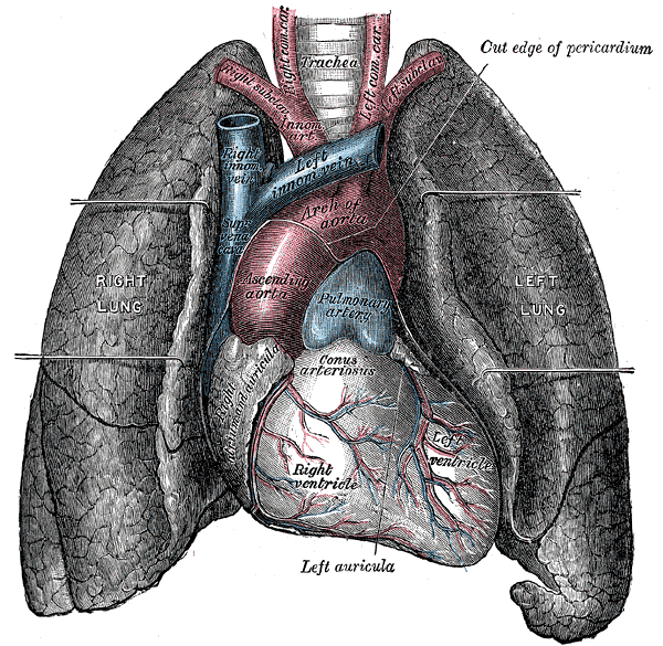
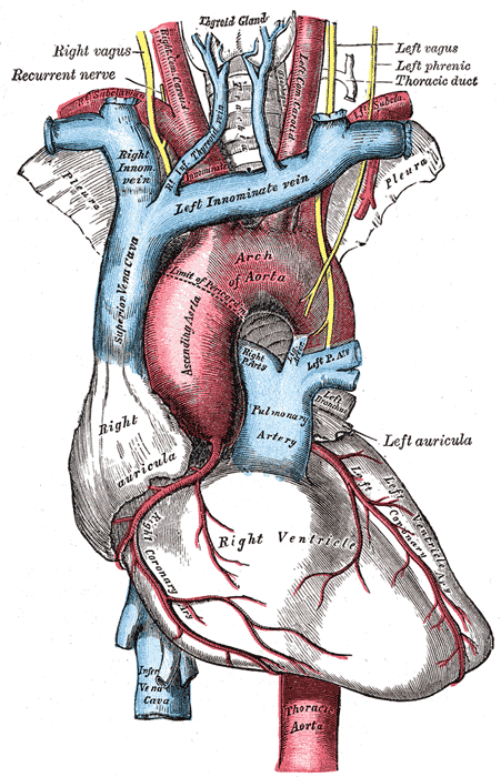
The heart and great vessels in situ, seen from the front with the lungs retracted.

Cardiovascular system / 7 min read

Heart

A complete guide to the human heart: chambers, valves and coronary vessels, the cardiac cycle, excitation-contraction coupling, cardiomyocyte biology, and the histology of all three cardiac layers.

Open study
02 / 15
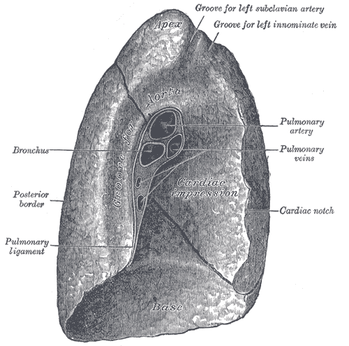
Mediastinal surface of the left lung, showing the hilum and cardiac impression.

Respiratory system / 6 min read

Lungs

How the lungs are built and how they work: the bronchial tree, pleura and respiratory mechanics, gas exchange and oxygen transport, alveolar cell biology, and the histology of the respiratory membrane.

Open study
03 / 15
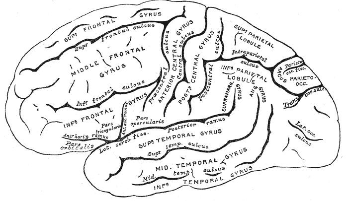
Lateral surface of the cerebral hemisphere, with the principal gyri and sulci named.

Nervous system / 6 min read

Brain

A structured guide to the human brain: lobes and deep structures, the meninges and cerebrospinal fluid, synaptic transmission, neuronal and glial biology, and the histology of grey and white matter.

Open study
04 / 15
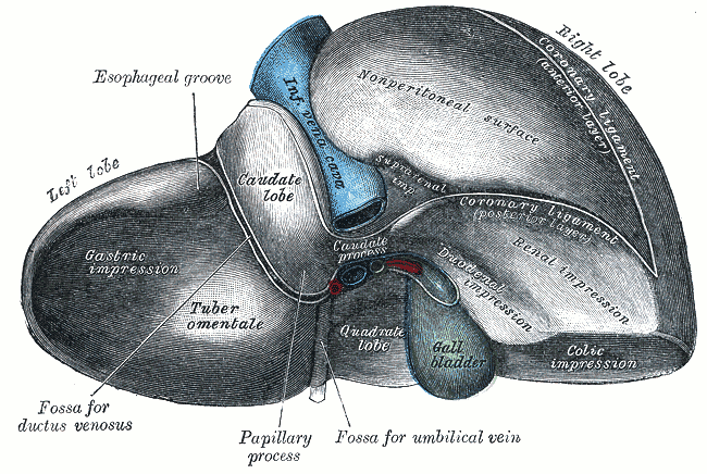
Visceral surface of the liver, showing the lobes, gallbladder and porta hepatis.

Digestive system / 7 min read

Liver

The liver explained in depth: lobes and segments, dual blood supply, metabolic and detoxification physiology, hepatocyte biology, the classic lobule and acinus, and the pathology of cirrhosis.

Open study
05 / 15
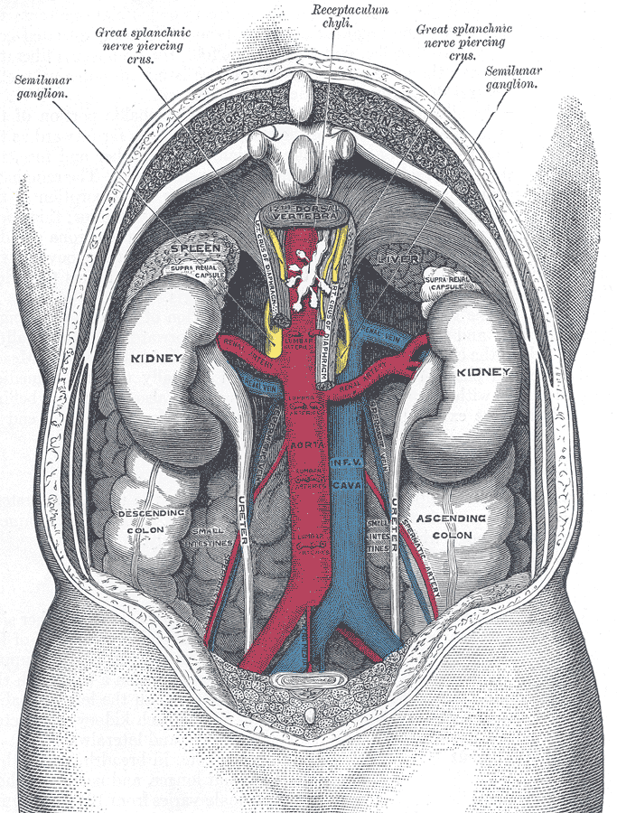
The kidneys and great vessels in situ on the posterior abdominal wall.

Urinary system / 7 min read

Kidneys

Follow blood through the nephron: renal anatomy, glomerular filtration, tubular transport, the countercurrent multiplier, hormonal control of volume and pressure, and renal histology segment by segment.

Open study
06 / 15
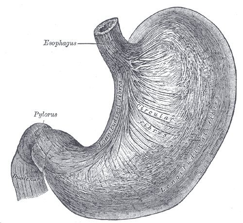
The stomach seen from the front, from the cardia through the body to the pylorus.

Digestive system / 7 min read

Stomach

How the stomach works: regions and wall layers, acid secretion by parietal cells, the three phases of gastric secretion, gastric gland biology, and the mucosal barrier that stops the organ digesting itself.

Open study
See all 15 organ guides

Tissues and building blocks / 10 studies

Start at the tissue.
The organ follows.

The layer underneath the organs: the four basic tissues, the cells they are built from, and how to tell them apart on a slide.
01 / 10
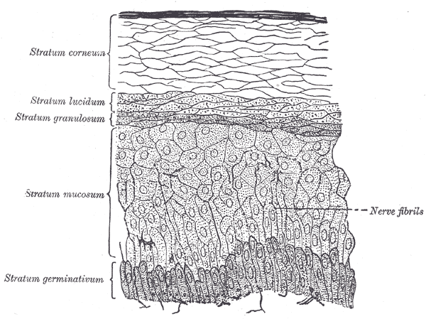
Section of the epidermis, showing the stacked layers of a stratified squamous epithelium.

Tissue biology / 9 min read

Epithelium

Every epithelium is named by two things: how many layers it has and what shape its surface cells are. A guide to simple, stratified, pseudostratified and transitional epithelium, what each one does, and how to tell them apart on a slide.
Open study
02 / 10
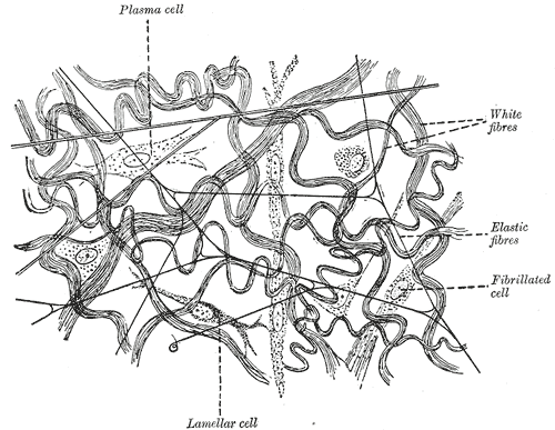
Loose subcutaneous tissue, highly magnified: collagen and elastic fibres scattered through ground substance.

Tissue biology / 9 min read

Connective tissue

Connective tissue is defined by what lies between its cells, not by the cells themselves. A guide to loose and dense connective tissue proper, the specialised forms — cartilage, bone, blood — and the matrix that gives each one its properties.

Open study
03 / 10
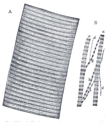
A human muscle fibre magnified nearly 800 diameters, with the myofibrils teased apart below.

Tissue biology / 9 min read

Muscle tissue

All three muscle types shorten by sliding the same two proteins past each other. What separates them is control, architecture and speed — and three features that let you tell them apart on a slide in seconds.

Open study
04 / 10
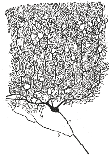
A cerebellar Purkinje cell after Cajal, showing the axon, the collateral, and the dendritic tree.

Tissue biology / 10 min read

Nervous tissue

Nervous tissue is two populations doing very different jobs: neurons, which carry signals and barely divide, and glia, which outnumber them, support them, insulate them, and give rise to most brain tumours.

Open study
05 / 10
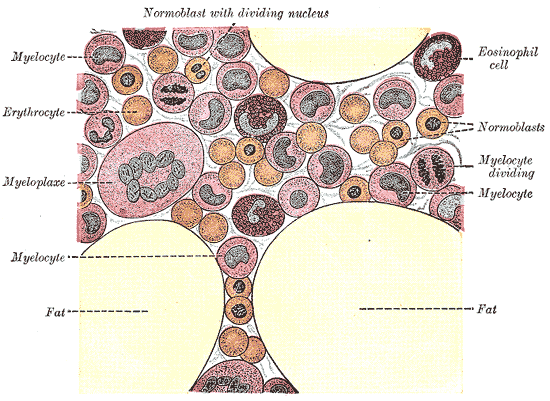
Human bone marrow, highly magnified — the tissue in which every blood cell is made.

Tissue biology / 10 min read

Blood

Blood is a connective tissue with a liquid matrix. What each of its four components does, where they are made, and what a full blood count is actually measuring.

Open study
06 / 10
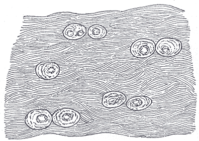
White fibrocartilage from an intervertebral disc, with chondrocytes set between dense collagen bundles.

Skeletal system / 9 min read

Cartilage

Three cartilages, three jobs, three appearances on a slide. Where each one is found, why none of them has a blood supply, and why that single fact explains almost every cartilage injury.

Open study
See all 10 tissue guides

Emergency medicine / 5 studies

Know the signs.
Act on the clock.

The time-critical conditions: how to recognise them, why minutes change the outcome, and what to do before help arrives.
A better way to look

From the whole organ to the living cell.

A body is not a collection of flashcards. Each Surgeonring study moves through four lenses in the same order, so that structure explains function instead of sitting beside it. The emergency guides follow the same order under plainer names: recognise it, understand why minutes matter, then know what to do.

  • Anatomy

    Where the organ sits, what it is attached to, and which vessels and nerves reach it.

  • Physiology

    The mechanism: what the organ actually does, step by step, and what regulates it.

  • Biology

    The cells that do the work, and why each one is shaped for the job it holds.

  • Histology

    What the tissue looks like under magnification, and how to recognise it on a slide.